


As humans, we are primarily active during the day and sleep at night. Because of that, our eyes are structured in such a way that our vision is clear when there is light (thanks to cone cells which we discussed in the previous article). Yet, during the night, we still can see, thanks to photoreceptor cells known as rod cells (rods). These cells are responsible primarily for low-light vision, known as scotopic vision (1). Because of their similarity to cones, both cell types are often discussed together.
At the back of the eye
Rod cells are found in the retina of the eye, in the same place as cones, although rods are usually present on the peripheries of the retina, while cones are located in the center. On average, there are 20 times more rods than cones (2). Both photoreceptor cells are elongated and slender, and can be divided into 2 segments: inner (that forms a synaptic terminal at one end of the cell) and outer (containing visual pigment) (2). The outer segment is, in fact, a highly modified cilium — an organelle formed by membrane projection, in this case by large amounts of lipid membranes (3). These membranes form invaginations (in cones), or discs (in rods), and store the visual pigment. To avoid the extensive growth of outer segments, new discs in rods are produced under the old ones, which are then degraded by retinal pigment epithelium (RPE) cells (4).
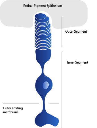
Inside the retina, rods and cones are located backward of the eye opening (with the outer segment located closer to the retina). There are a few reasons for that. Firstly, such a situation allows proper access to the outer segment for RPE cells, which is needed for phagocytosis, but also for providing nutrients back to rods and cones. Secondly, RPE are absorbing the majority of scattered light, which saves rod and cone cells from unnecessary light damage (1).
The Dark Side of The Moon
Another difference between cones and rods is their sensitivity to light. Rod cells can detect as little as one photon of light, while cone cells require hundreds of times more to be activated (5). At the same time, cone cell activation is much faster, and provides higher resolution. Cones are also better at adapting to brightness (5). Rod cells’ ability to be activated by so little light allows us to see the shapes and contours of objects at night. Interestingly, rods get saturated by even moderate amounts of light; therefore they remain inactive during the day (6).
The type of visual pigment can also affect vision sensitivity. There are two types of pigments: opsins in cones (absorbing blue, green, or red light) and rhodopsin in rods (which can absorb light of a shorter wavelength than opsins). Both opsins and rhodopsin belong to G-protein coupled receptor (GPCR) family, proteins that are embedded in the membrane to be activated by their ligand. Unlike other GPCRs, the ligand of rhodopsin (called retinoid ligand) is pre-bound to it so that it can be activated upon binding of a photon (2).
Night shift mode
We live in a day-and-night cycle. To get used to this rhythm, our retinas are utilising a complex circuit that is switched ON and OFF depending on the presence of the light (7). When the light is gone, the circuit is taken over by the rods (7).
Such night shifts can be however easily disturbed by the presence of short-wavelength blue light. Many electronic devices emit this type of light. That’s why it is recommended not to use smartphones or computers in the evening since their light can affect our sleep, which in the long run can lead to the development of mental diseases such as depression (8).
On the other hand, it was suggested that blue light emitted by electronic devices can cause age-related macular degeneration (AMD), and eye disease leading to blurring of the central vision. However, a recent study on a large group of people who were using blue light filters while working shows that there is no link between the two factors (9). In another study, it was shown that a lack of rod cells decreases the development of retinal neovascularisation (growth of new abnormal blood vessels, caused by eye injuries or diseases) while suppressing rods using blue light at night decreases the rate of progression of early diabetic retinopathy (10). However, a 24-month-long study showed that limiting light at night by wearing eye masks by patients with diabetic macular oedema had no therapeutic benefit (11).
Recognizing and appreciating the labs working in this space
- Krzysztof Palczewski Lab, Center for Translational Vision Research, University of California, USA https://faculty.sites.uci.edu/palczewskilab/
- AZ Retina Project — azretina.sites.arizona.edu , Twitter: @AZretinaproject
- Humberto Fernandes Lab, ICTER, Poland https://icter.pl/research/integrated-structural-biology/ , Twitter: @ICTER_PL , Instagram: https://www.instagram.com/icter_research/ , Facebook: https://www.facebook.com/icterresearch
- Arendt Group, EMBL Heidelberg, https://www.embl.org/groups/arendt/
- Stenkamp Lab, University of Idaho, USA, https://www.stenkamplab.net/
References
- Kazilek, CJ, and Cooper, Kim. “Rods and Cones.” ASU — Ask A Biologist. (2010). https://askabiologist.asu.edu/rods-and-cones
- Hofmann, Klaus Peter, and Trevor D Lamb. “Rhodopsin, light-sensor of vision.” Progress in retinal and eye research vol. 93 (2023): 101116. doi:10.1016/j.preteyeres.2022.101116
- May-Simera, Helen et al. “Cilia — The sensory antennae in the eye.” Progress in retinal and eye research vol. 60 (2017): 144–180. doi:10.1016/j.preteyeres.2017.05.001
- Mazzoni, Francesca et al. “Understanding photoreceptor outer segment phagocytosis: use and utility of RPE cells in culture.” Experimental eye research vol. 126 (2014): 51–60. doi:10.1016/j.exer.2014.01.010
- Krishnamoorthi, Arjun et al. “Ultrafast Transient Absorption Spectra and Kinetics of Rod and Cone Visual Pigments.” Molecules (Basel, Switzerland) vol. 28,15 5829. 2 Aug. 2023, doi:10.3390/molecules28155829
- Green, D G. “Light adaptation in the rat retina: evidence for two receptor mechanisms.” Science (New York, N.Y.) vol. 174,4009 (1971): 598–600. doi:10.1126/science.174.4009.598
- Sivyer, Benjamin, and Henrique von Gersdorff. “Vision: Microcircuits Rage against the Dimming of the Light.” Current biology : CB vol. 28,20 (2018): 3353. doi:10.1016/j.cub.2018.10.020
- Newsom, Rob, and Singh, Abhinav. “How Blue Light Affects Sleep.” Sleep Foundation (2023). https://www.sleepfoundation.org/bedroom-environment/blue-light
- Mainster, Martin A et al. “The Blue Light Hazard Versus Blue Light Hype.” American journal of ophthalmology vol. 240 (2022): 51–57. doi:10.1016/j.ajo.2022.02.016
- Sivaprasad, S, and G Arden. “Spare the rods and spoil the retina: revisited.” Eye (London, England) vol. 30,2 (2016): 189–92. doi:10.1038/eye.2015.254
- Sivaprasad, Sobha et al. “Clinical efficacy and safety of a light mask for prevention of dark adaptation in treating and preventing progression of early diabetic macular oedema at 24 months (CLEOPATRA): a multicentre, phase 3, randomised controlled trial.” The lancet. Diabetes & endocrinology vol. 6,5 (2018): 382–391. doi:10.1016/S2213–8587(18)30036–6
About the author:
DR. MAŁGORZATA ‘MASIA’ MAKSYMOWICZ
Content Editor The League of Extraordinary Cell Types, Sci-Illustrate Stories
Dr. Maksymowicz did her Ph.D. in Cell Biology (IIMCB, Poland) studying the intracellular trafficking and inflammatory signalling of a cytokine receptor. She did a 1-year post-doc at Nencki Institute, Poland, studying the protein- and RNA-binding properties of proteins. Currently, she is doing a post-doc at Barts Cancer Institute, UK, studying the links between endocytosis and tumorigenesis. Dr. Maksymowicz is passionate about science and loves to combine different fields of biology, always trying to seek beauty in nature.
About the artist:
NELLY AGHEKYAN
Contributing Artist The League of Extraordinary Cell Types, Sci-Illustrate Stories
Nelli Aghekyan, did a bachelor’s and master’s in Architecture in Armenia, after studying architecture and interior design for 6 years, she concentrated on her drawing skills and continued her path in the illustration world. She works mainly on children’s book illustrations, some of her books are now being published. Currently living in Italy, she works as a full-time freelance artist, collaborating with different companies and clients.
About the animator:
DR. EMANUELE PETRETTO
Animator The League of Extraordinary Cell Types, Sci-Illustrate Stories
Dr. Petretto received his Ph.D. in Biochemistry at the University of Fribourg, Switzerland, focusing on the behavior of matter at nanoscopic scales and the stability of colloidal systems. Using molecular dynamics simulations, he explored the delicate interaction among particles, interfaces, and solvents.
Currently, he is fully pursuing another delicate interaction: the intricate interplay between art and science. Through data visualization, motion design, and games, he wants to show the wonders of the complexity surrounding us.
About the series:
The League of Extraordinary Cell types
The team at Sci-Illustrate and Endosymbiont bring to you an exciting series where we dive deep into the wondrous cell types in our body, that make our hearts tick ❤.